Multidisciplinary Treatment of a Patient
with Craniofacial Disorders
Interdisciplinary care typically begins with the general dentist, as does the patient’s belief in the possibilities of treatment. Development
of trust and cohesion among dental team members is avital benefit of belonging to study clubs. The following multidisciplinary case illustrates not only an excellent working relationship among dental professionals, but a shared determination of common treatment goals, which, in turn, developed the faith and confidence needed from the patient to proceed with treatment
Diagnosis
A 36-year-old male presented to Dr. Thompson’s office after having seen multiple dentists, all of whom had provided little but palliative care. Craniofacial distortions, poor oral hygiene, and speech difficulties seemed to have convinced previous professionals that he was unwilling to participate in his care. Dr. Thompson assured the patient that with good cooperation, his mouth could be restored to a healthy state, after which he could be helped to obtain a natural smile that he could be proud of. From thatmoment on, if there were any deficiencies in his care, they did not result from any lack of participation with his dental team.
The patient’s medical history was unremarkable except for an isolated cleft of the soft palate, for which he had undergone two surgical closure procedures. He also showed indications of a failed posterior pharyngeal flap.
The clinical examination revealed a Class II malocclusion, severe crowding, multiple decayed teeth, impactions, a missing lower left second molar, and an extreme anterior open bite with dental protrusion (Fig. 1).
Fig. 1 36-year-old male with multiple craniofacial distortions, chronic marginal gingivitis, Class II highangle skeletal pattern with constricted dental arches, posterior dentoalveolar eruption, excessive anterior facial height, recessive mandible, and severe anterior open bite before treatment.
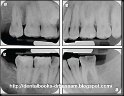
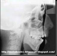
Chronic marginal gingivitis was evident, with greater than 50% bone loss on the labial aspects of both maxillary canines. Radiography confirmed the Class II malocclusion and showed a high-angle skeletal pattern with constricted dental arches, posterior dentoalveolar eruption, excessive anterior facial height, a recessive mandible, and an anterior open bite.
Treatment Plan
Dr. Thompson consulted with an orthodontist (Dr. Mehan) and an oral surgeon (Dr. Hochberg) to develop an appropriate multidisciplinary treatment plan, which was presented to the patient for approval. The agreedupon treatment sequence was as
follows:
1. Treatment of carious teeth, periodontal control, and monitoring of home care.
2. Surgical removal of both maxillarycanines, both mandibular second premolars, and the maxillary third molars; uncovering of the impacted maxillary right second premolar; and placement of skeletal anchors cervical to the mandibular first molars.
3. Orthodontic treatment involving the use of self-ligating appliances and light-wire forces to relieve crowding and align the dentition; intrusion of the mandibular posterior teeth with skeletal anchorage; placement of a lingual arch to prevent buccal rotation of the mandibular first molars; light elastic wear; and, finally, placement of vacuumformed retainers.
4. Evaluation of restorative needs, including vital bleaching and permanent restorations.
5. Plastic surgery.
6. Speech evaluation, in comparison with recordings made before orthodontic care, to determine the need for reestablishment of a posterior pharyngeal flap that would allow the production of normal nasal sounds.
Treatment Progress
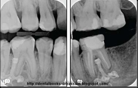
After treatment of the dental caries and stabilization of the soft-tissue inflammation, the maxillary canines, mandibular second premolars, and maxillary third molars were extracted. The impacted maxillary right second premolar was surgically uncovered, and titanium miniplates** were placed with three 4mm monocortical miniscrews in the right and left mandibular buccal cortices, approximately 8mm cervical to the cemento-enamel junctionsof the first molars. Intrusive forces were applied with elastic thread from the protruding portions of the miniplates to the first molar brackets. A mandibular lingual arch was placed to prevent buccal rotation during intrusion.
Full orthodontic appliances were placed seven months after the start of treatment (Fig. 2)
Fig. 2 Eight weeks after placement of brackets and mandibular lingual arch.
and orthodontic treatment proceeded uneventfully for 17 months (Fig.3A).
(Fig.3A).
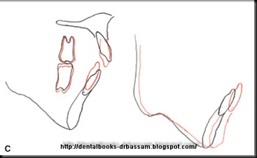
The bite was closed both dentally, through the retraction of anterior teeth, and skeletally, by intrusion of the mandibular buccal segments and autorotation of the mandible (Fig. 3C).
Fig. 3C
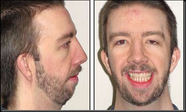
Further planned treatments include facial plastic surgeries; prosthetic replacement of the missing lower left second molar; porcelain/ceramic crowns for the upper left second molar, lower left first molar, and lower right canine and second molar; and soft-tissue grafts for the upper left central incisor, upper left first molar, and lower left canine. Although the surgical and restorative procedures have not yet been completed, the esthetic improvement already achieved is remarkable (Fig. 4).
Fig. 4 A. Patient after 27 months of multidisciplinary treatment. Vertical position of upper left second molar will be maintained with vacuum-formed retainers until future prosthetic replacement of lower left second molar. B. Miniplates left in place after end of treatment for anchorage in case of relapse (none seen to date).
Comparison of the preand post-treatment cephalometric radiographs shows a considerable reduction in facial height and an increase in the projection of the lower facial third. An advancement genioplasty would be helpful at some point, but the improvement in the patient’s smilehas already changed his life.
Conclusion
The significance of this case report is not in how the patient was treated—many practitioners could have been equally successful.
It was the trust established between the patient and the interdisciplinary team that made this enhanced treatment possible in the first place. We may be surprised how positively people will respond when we show a sincere interest in them.
WILLIAM A. MEHAN, DMD, MS
PAUL THOMPSON, DDS
MARK HOCHBERG, DMD





No comments:
Post a Comment